Аналізатори газів крові та електролітів
")
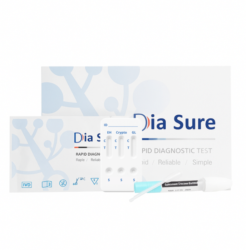
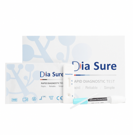
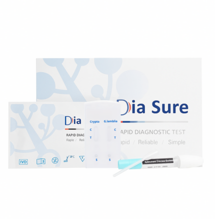
Швидкий комбі-тест для визначення E. histolytica, Cryptosporidium та Giardia lamblia в калі
Високочутливий імунохроматографічний експрес-тест in vitro, призначений для якісного виявлення Entamoeba histolytica, Cryptosporidium spp. та Giardia lamblia у зразках калу людини.
Простота використання, швидке отримання результату (протягом 10 хвилин) та відсутність потреби у спеціальному лабораторному обладнанні роблять цей тест-набір ефективним інструментом для початкової діагностики амебіазу, криптоспоридіозу та лямбліозу в умовах лікарень, клінік, лабораторій і польових точок тестування. Тест забезпечує високу точність завдяки специфічному виявленню антигенів трьох основних паразитарних інфекцій із чутливістю понад 97% та специфічністю понад 99%.
Отримані результати слід інтерпретувати у поєднанні з клінічними симптомами та іншими лабораторними методами у разі потреби.
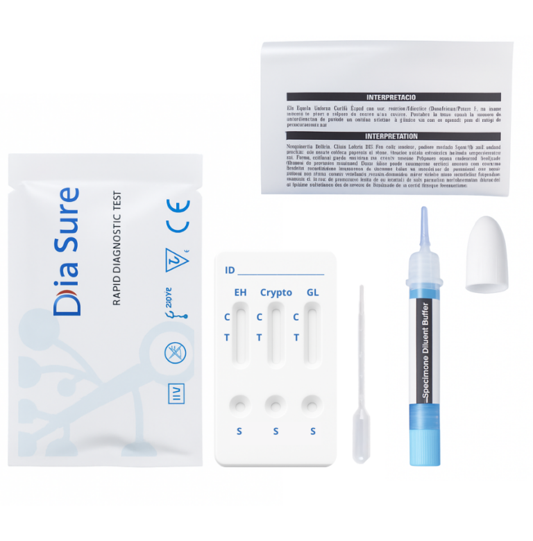
КОМПЛЕКТАЦІЯ ТЕСТ-НАБОРУ
Що входить у комплект?
Індивідуально упаковані прилади для тестування (касети)
Пакет-вкладиш (інструкція)
Одноразові піпетки
Пробірка для розведення зразків з буфером
ПРОЦЕДУРА ДОСЛІДЖЕННЯ

Перед використанням доведіть тести, зразки, буфер та/або контрольні зразки до кімнатної температури (15–30°C).
Збір зразків і підготовка
Використовуйте пробірку для збору та розведення зразків, що входить у комплект. Дотримуйтесь інструкції на упаковці. Також можна використовувати інші чисті та сухі контейнери. Найкращі результати досягаються, якщо аналіз проводиться протягом 2 годин після збору.
Для твердих зразків: відкрутіть аплікатор пробірки з буфером. Обережно, не пролийте вміст. Вставте аплікатор у щонайменше 3 різні ділянки калу, щоб зібрати приблизно 120 мг.
Для рідких зразків: тримайте піпетку вертикально, наберіть зразок і перенесіть 3 краплі (приблизно 120 мкл) у пробірку з буфером.
Поверніть аплікатор у пробірку, щільно закрийте кришку. Не зламайте наконечник. Добре струсіть пробірку, щоб зразок повністю змішався з буфером.
Зразки можна зберігати при+2…+8°C до 3 днів.
Проведення тесту
1. Вийміть тест з упаковки та розмістіть на чистій, рівній поверхні. Позначте тест (пацієнт або контроль).
2. Зламайте наконечник пробірки (наприклад, за допомогою серветки). Тримайте вертикально, нанесіть 2 краплі у лунку для зразка (S). Уникайте бульбашок повітря. Не капайте в оглядове вікно.
3. Після запуску тесту колір почне рухатись мембраною. Зачекайте 10 хвилин. Результати, зчитані після 20 хвилин, вважаються недійсними.
ІНТЕРПРЕТАЦІЯ РЕЗУЛЬТАТІВ

ПОЗИТИВНИЙ
На мембрані з'являються дві кольорові смуги. Одна смуга з'являється в контрольній області (C), а інша — в тестовій області (T).

НЕГАТИВНИЙ
У контрольній області (C) з’являється лише одна кольорова смуга. У тестовій області (T) кольорова смуга не з’являється.